
F Waves Ekg
The normal U wave has the same polarity as the T wave and is usually less than one-third the amplitude of the T wave.
F waves ekg. The QRS complex, normally beginning with a downward deflection, Q;. A 60-year-old female asked:. (1)Feinberg School of Medicine, Northwestern University, USA.
The f wave is modified by the near field potential and the far field potential. Traversal of F-waves along the entire length of periphera. Anthony Kashou (The EKG Guy) is a physician resident at the Mayo Clinic in Rochester, Minnesota.
See detailed information below for a list of 4 causes of Absent P waves on ECG, Symptom Checker, including diseases and drug side effect causes. He is on a mission to transform ECG education and filling the gap exists around the world. That doesn’t just mean standing up to high-winds and hail, or whatever else nature throws down.
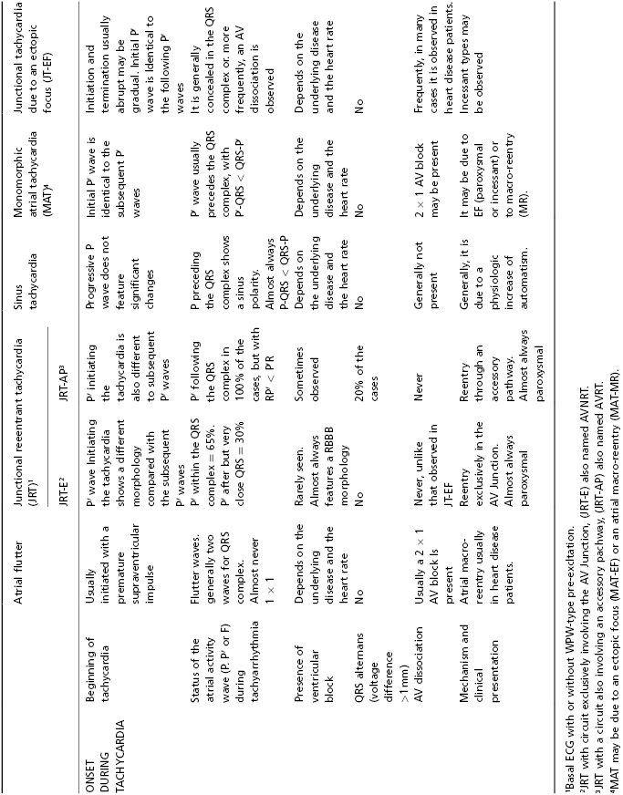
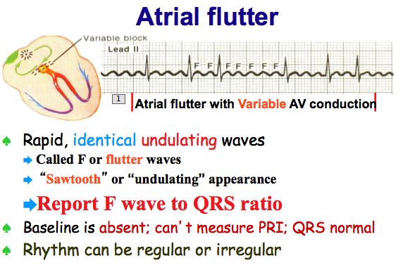
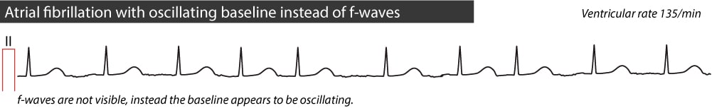
The amplitude of f wave arises along with the deepening of the catheter tip. 1st negative deflection of QRS complex after P wave or before 1st R wave ;. Atrial flutter is the only diagnosis causing this baseline appearance, which is why it must be recognized on the ECG.
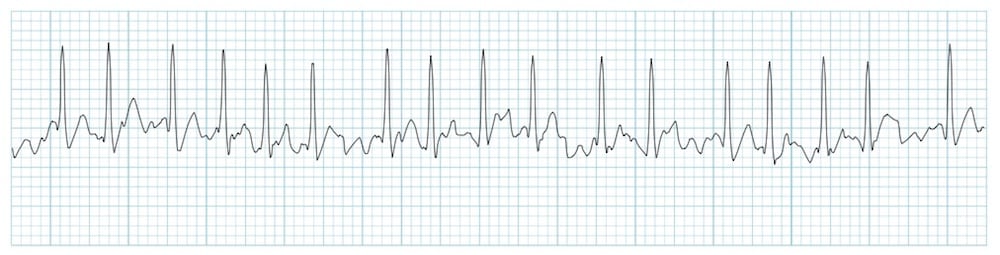
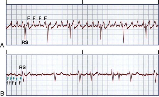
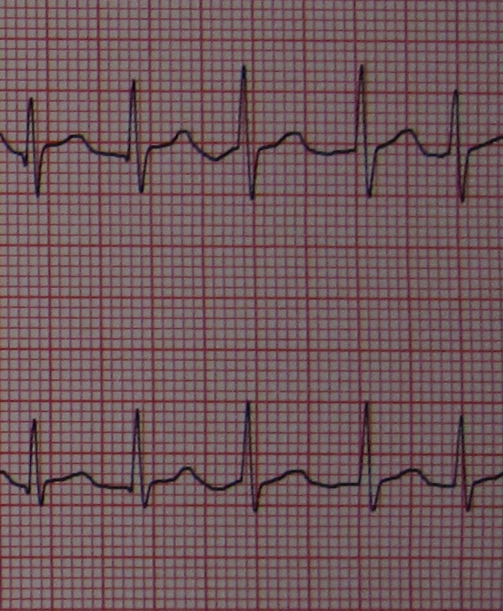
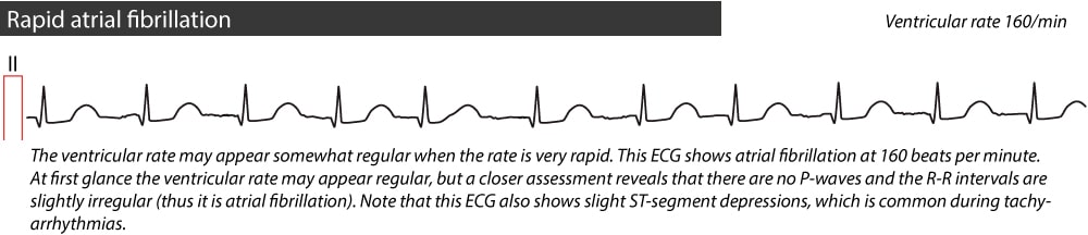
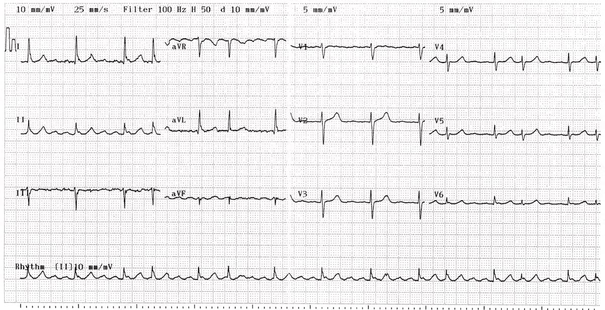
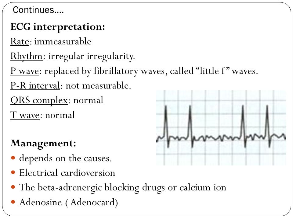
The baseline (isoelectric line between QRS complexes) is characterized by either fibrillatory waves (f-waves) or just minute oscillations. Less commonly, F waves are undulating, firing at a rate of 280-3/min, a rate often associated with a 2:1 block and alternating F waves merge with QRS or T waves Neurology. Electrocardiography is the process of producing an electrocardiogram (ECG or EKG).It is a graph of voltage versus time of the electrical activity of the heart using electrodes placed on the skin.
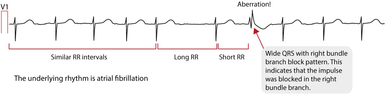
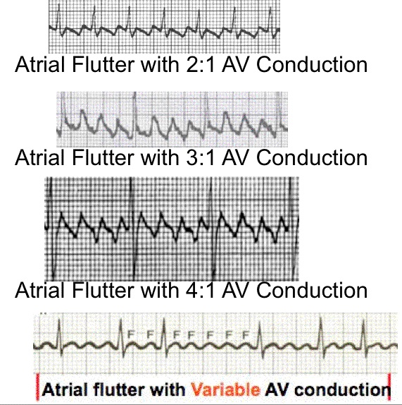
Not to be confused with f-waves seen in atrial fibrillation) which gives the baseline a saw-tooth appearance. The R-R intervals are not equal resulting in an irregular rhythm (irregularly irregular). Absence of pathologic Q waves does not exclude a myocardial infarction!.
Inverted T waves are normal in children, and they sometimes remain inverted into adulthood. The different waves that comprise the ECG represent the sequence of depolarization and repolarization of the atria and ventricles. What do "slightly flat" s/t waves mean on a routine ekg?.
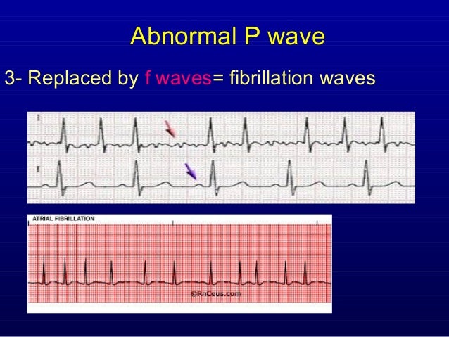
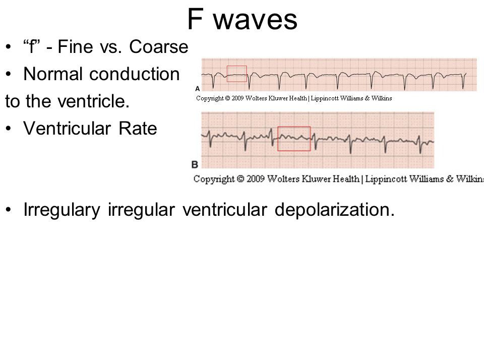
The P wave should be upright in lead II if the action potential is originating from the SA node. This chaotic electrical activity results in a chaotic wave form between the QRS complexes. (A lowercase f indicates atrial fibrillation).
Waves are the different upward or downward deflections represented on the EKG tracing. The ECG shows regular flutter waves (F-waves;. An electrocardiogram (ECG or EKG) records the electrical signal from your heart to check for different heart conditions.
When the tip of PICC comes to the ideal position (the lower 1/3 of SVC), f wave amplitude is the highest, which is similar to the change of P wave in the normal surface ECG patients. On the horizontal axis, each large box represents 0.2 seconds, and each smaller box. It results from the backfiring of antidromically activated horn cells.
The PRI is indeterminate. Massage of the carotid sinus may unmask the flutter waves. Send thanks to the doctor.
At F Wave, we have one simple mission:. There are fibrillation (f) waves instead of P waves. Anthony Kashou (The EKG Guy) is a.
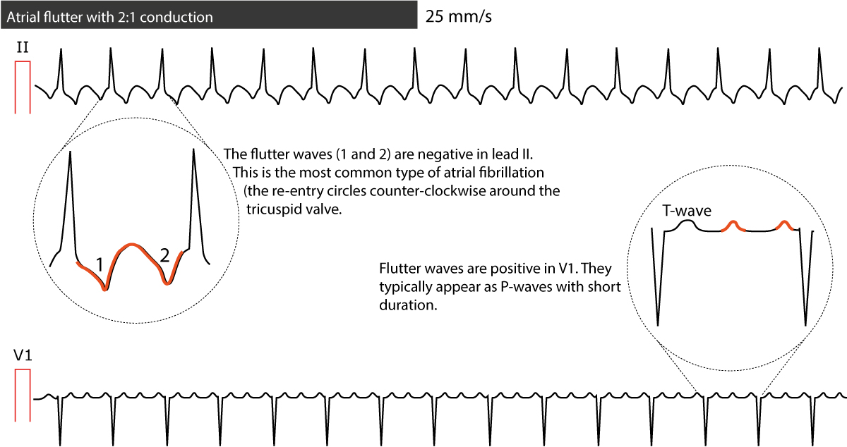
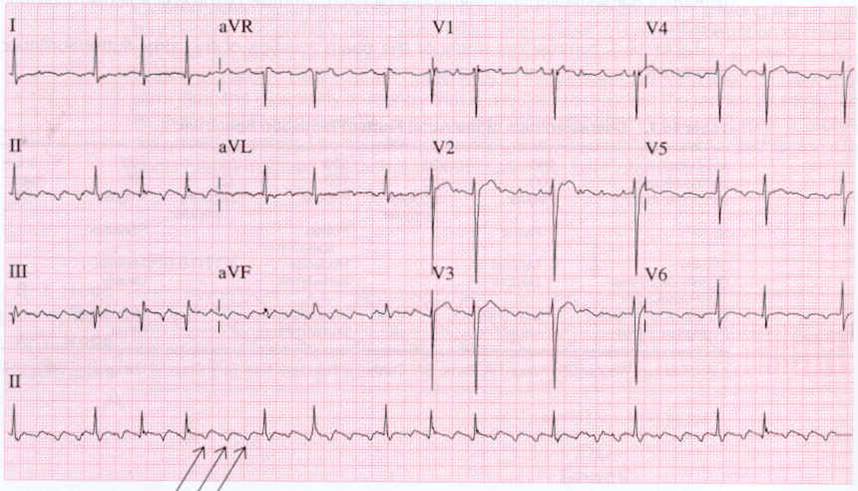
But lead V1 records definite isoelectric lines. Frequently the request is made by practitioners with no particular expertise in cardiology. Electrocardiogram waves, intervals, and segments.
Q waves of 0.04 seconds (1 mm) duration and greater than one third the R wave's amplitude in the same lead may be pathological. They are the product of the action potentials created during the cardiac stimulation, and repeated from one heart beat to another, barring alterations. Although heart rate can be calculated easily by taking a pulse, studies show that an ECG (electrocardiogram) may be necessary to determine if there is any damage to the heart, how well a device or drug is working, whether the heart is beating normally, or to determine the location and size of the heart chambers.
The first little “hump” or “bump” you see is known as the P-wave. (2)Vanderbilt University Medical Center, Nashville, Tennessee, USA. An electrocardiogram (ECG) may be requested as part of the investigation of a wide range of problems in paediatrics, often in patients who have no clinical evidence of cardiac disease.
Nakayama M, Nakamura J, Hamada Y, Chaya S, Mizubayashi R, Yasuda Y, Kamiya H, Koh N, Hotta N Diabetes Care 01 Jun;24(6):1093-8. Am J Emerg Med. Create the best shingle the world has ever seen.
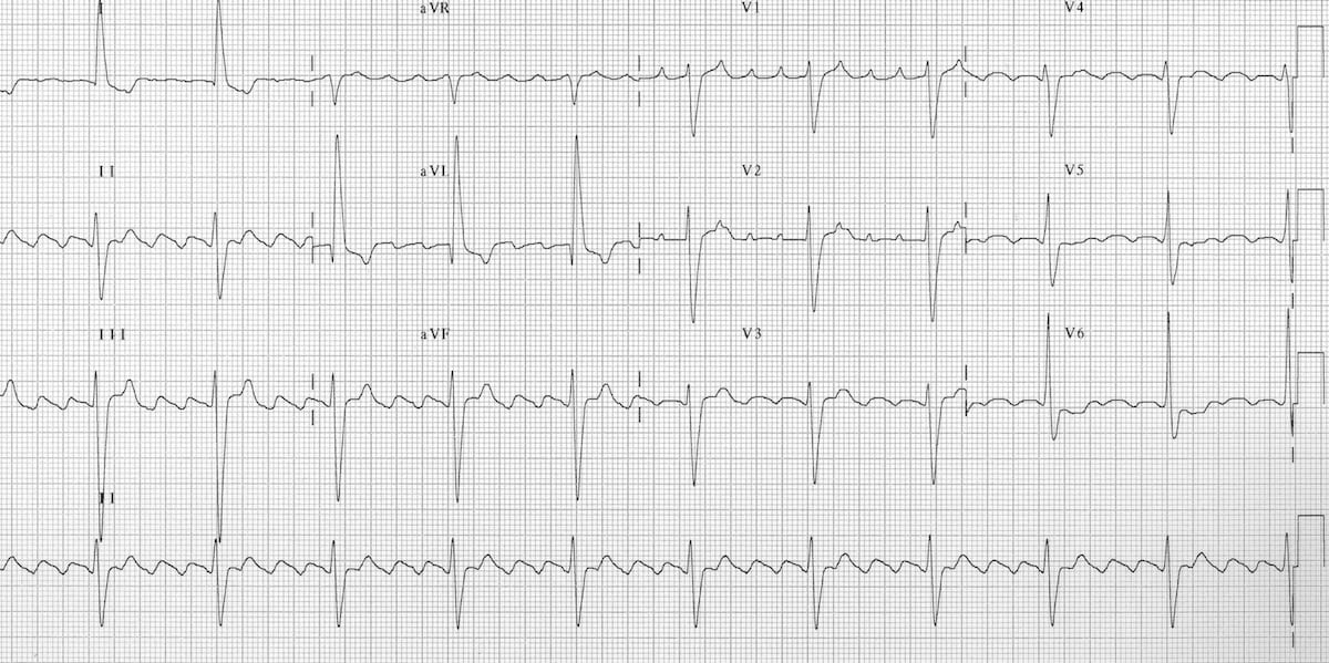
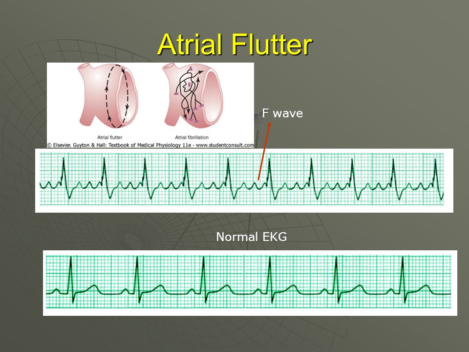
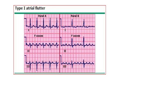
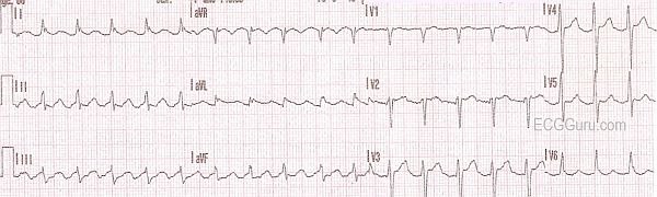
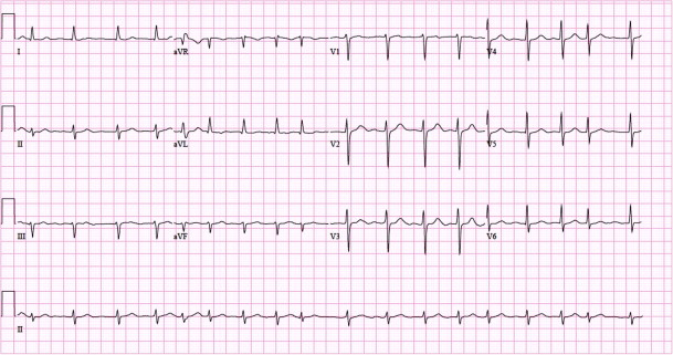
U waves are usually best seen in the right precordial leads especially V2 and V3. Cardiology An atrial flutter wave on EKG, which appears as a 'sawtooth' pattern in leads II, III and aVF;. These need to be present in at least 2 contiguous.
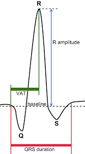
If we move along the graph of the ECG, we see a small dip followed by a large spike and another dip. Approximately 80% of the population is plagued at one time or another by back pain. Indeed, a repeat ECG (figure 1C) confirms the presence of P waves.
Some common causes of R wave abnormalities on an ECG include a thin chest wall or obesity. The atria quiver rapidly, with most electrical impulses being blocked before reaching the ventricles. A larger upwards deflection, a peak (R);.
A Q wave is any negative deflection that precedes an R wave. The F wave occurs after the direct motor potential or the M response. Lead III often shows Q waves, which are not pathologic as long as Q waves are absent in leads II and aVF (the contiguous leads).
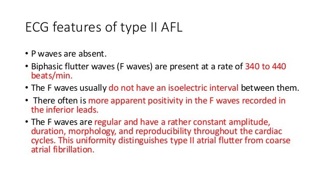
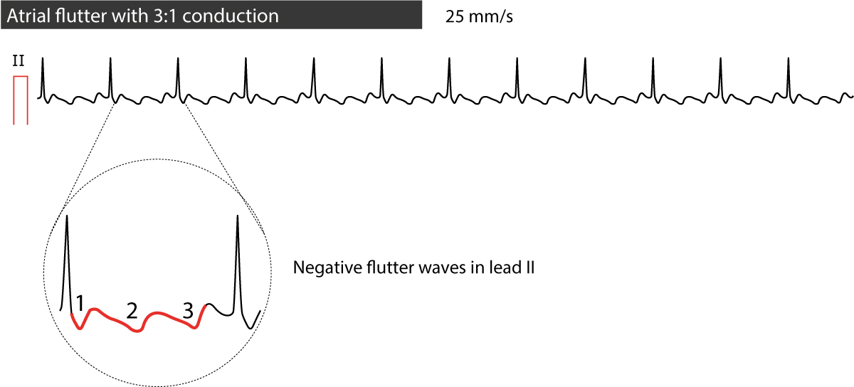
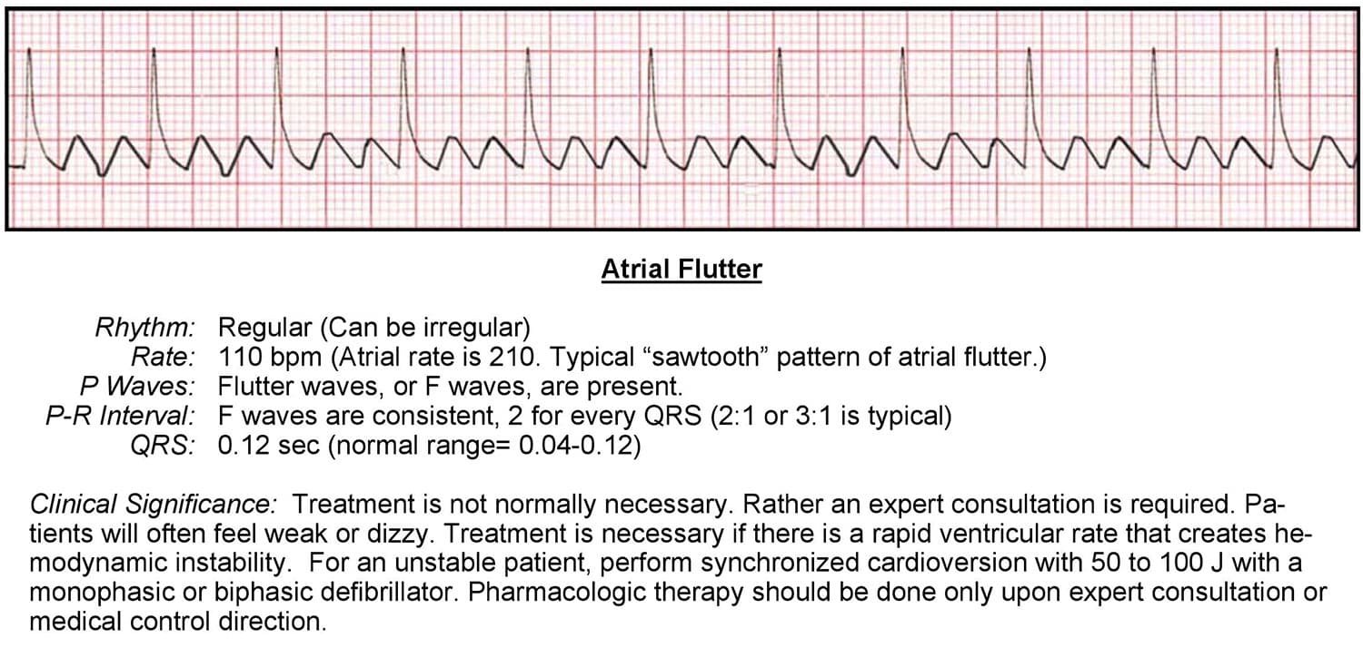
Kashou has taught and developed curriculum for medical students, including 500+ lectures and 100+ hours of adult and pediatric ECG lessons. • Flutter waves (F waves) are seen when the impulse arises from atria at a rate of 250 to 350 BPM. Sovari AA, Farokhi F, Kocheril AG.
R-wave ≥ 0.04 s in V1–V2 and R/S ≥ 1 with a concordant positive T-wave in the absence of a conduction defect. The P-wave represents ATRIAL DEPOLARIZATION (depolarization is a big, fancy word for CONTRACTION). Sharma P(1), Barrett TW(2), Ng J(3), Knoten C(3), Ferreira AJ(2), Goldberger JJ(3)(4).
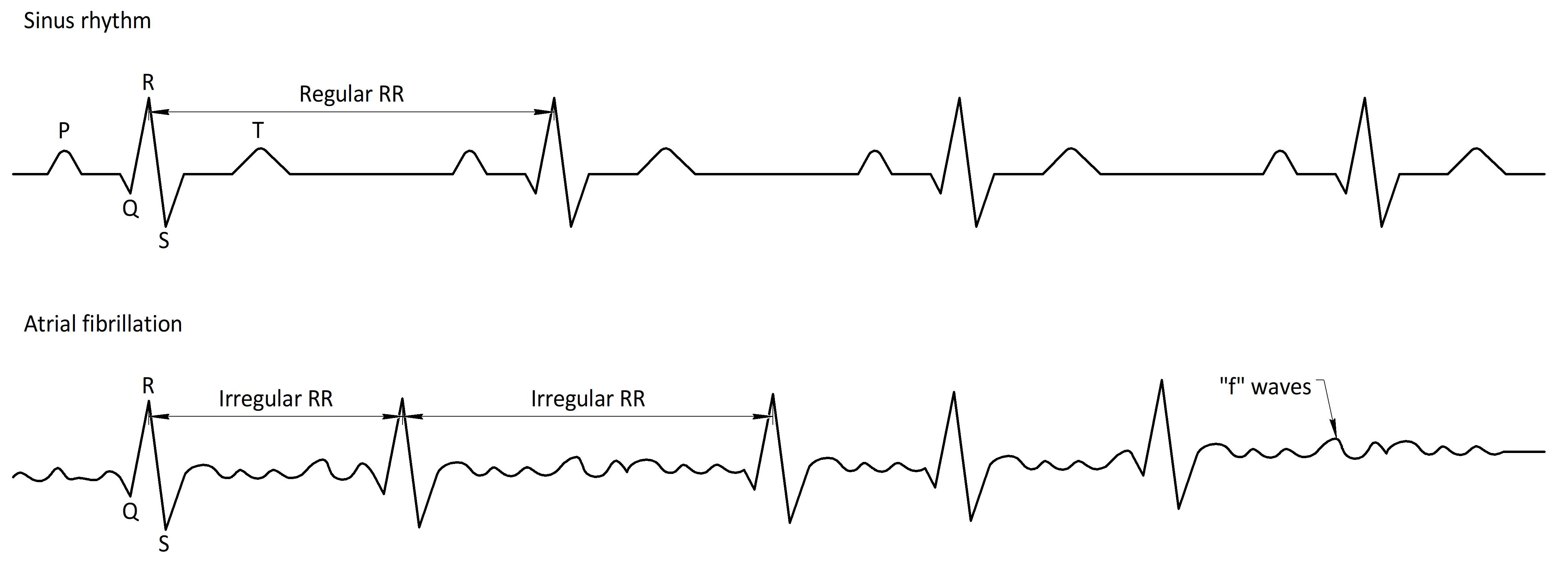
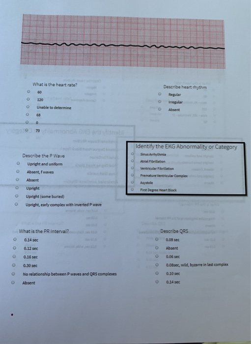
P waves are absent. It indicates that the atria are contracting, pumping blood into the ventricles. » Review Causes of Absent P waves on ECG:.
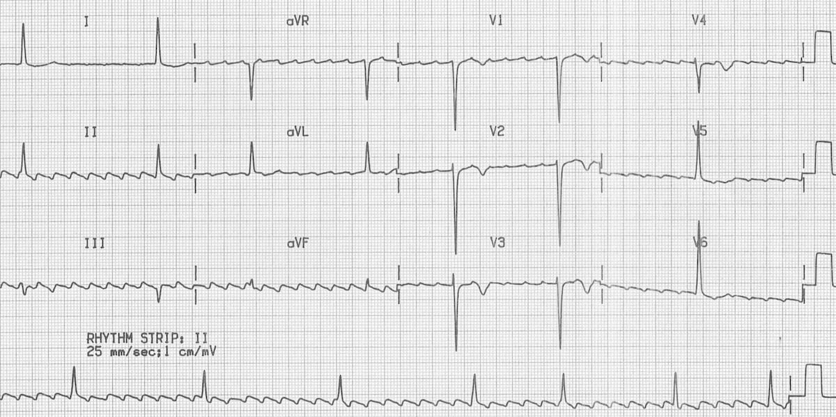
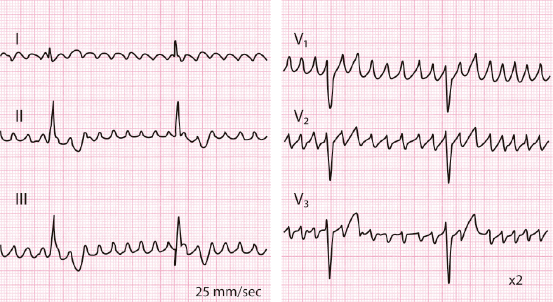
Cardiologists and other health professionals analyze ECGs to diagnose various cardiac diseases. If the rhythm is fast - (narrow complex tachycardia without P waves) Then it’s an SVT. The ECG is recorded at a speed of 25 mm/sec (5 large squares/sec), and the voltages are calibrated so that 1 mV = 10 mm (2 large squares) in the vertical direction.
If it’s like this - Then it’s a Junctional Rhythm. This test detects the electrical activity of the heartbeat through electrodes. The Q wave represents the normal left-to-right depolarisation of the interventricular septum;.
F response An undulation of an EMG that corresponds to the time between application of a stimulus to the axon of the α motor neuron as it propagates andromically to the anterior horn of the spinal cord, and. Causes | Symptom Checker » Causes of Absent P waves on ECG:. No P waves means there is no PR interval measurement.
Johnstrude CL, Perry JC, Cecchin F, et al. (supraventricular tachycardia) If the rhythm is like this - It’s “A-Fib” or atrial fibrillation. Absent P waves on ECG:.
And then a downwards S wave. Note the 'Flutter' or 'F' waves. It means looking great while doing it.
The basic principles of interpretation of the ECG in children are identical to those in adults, but the progressive. Clear 'flutter waves' can be seen. This is, in fact, a marked sinus arrhythmia, an ECG finding frequently mistaken for atrial fibrillation.
Flat t waves are a nonspecific finding, in and of themselves they are of minimal significance. P wave is the first short upward movement of the ECG tracing. Inverted U wave, a specific electrocardiographic sign of cardiac ischemia.
The normal U wave is asymmetric with the ascending limb moving more rapidly than the descending limb (just the opposite of the normal T wave). Remember from the electrical conduction lecture, that the SA node is responsible for this. The F-wave is a long latency muscle action potential seen after supramaximal stimulation to a nerve.
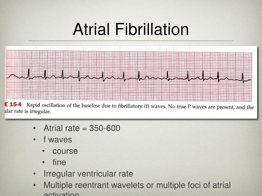
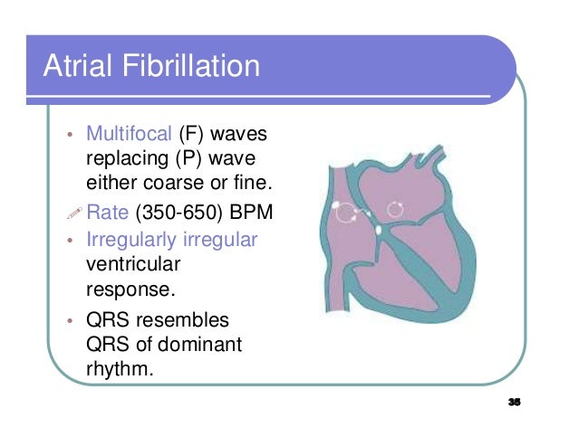
Atrial Fibrillation occurs when multiple electrical impulses occur within within the atria. This leads to an extremely high and unproductive atrial rate, but throttled ventricular rate. The F wave uses supramaximal stimulation of a motor nerve and records compound muscle action potentials from a muscle supplied by that nerve, along the most proximal segment.
In this setting, the ECG is said to demonstrate a normal sinus rhythm, or NSR. Inverted T waves may indicate several conditions, including pulmonary embolism, hypertrophic cardiomyopathy and heart attack. Atrial Flutter and Atrial Fibrillation With atrial flutter the 'P' waves are indeterminate.
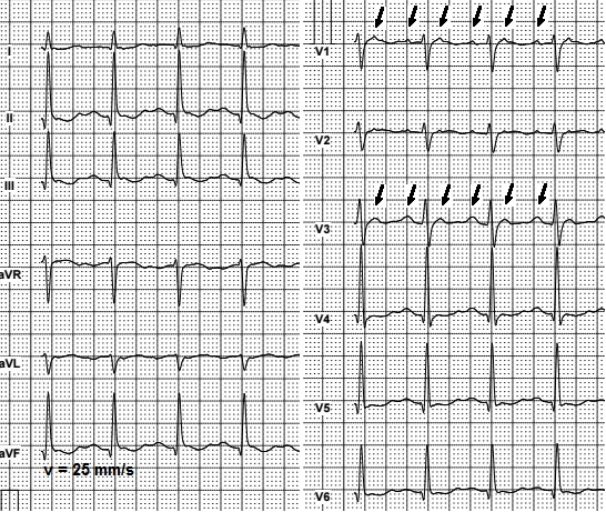
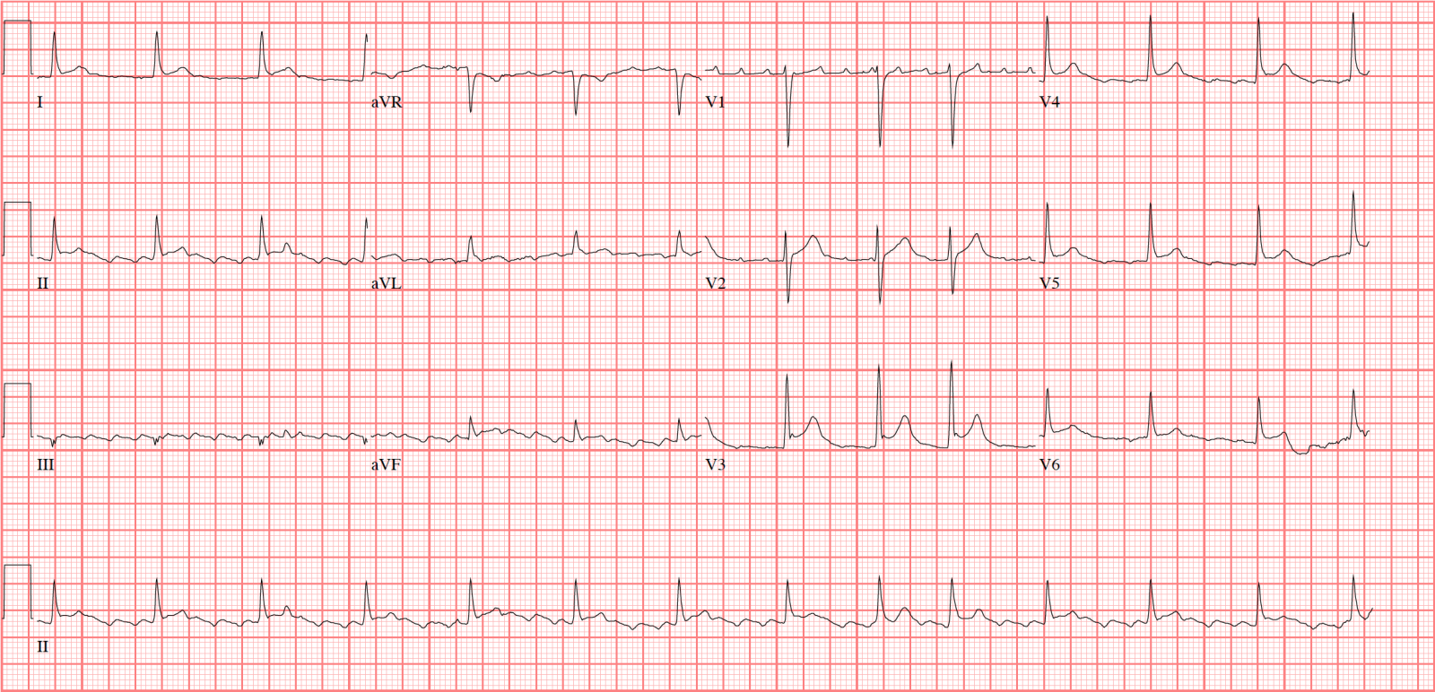
The pathological Q waves seen in V1 - V6 indicate that this patient has had an anterior MI in the past. On standard calibration, each large box has sides of 0.5 cm. Associated leg pain occurs less frequently.
This patient also has evidence of an acute inferior MI as shown by the ST segment elevation in leads III and aVF. An EKG readout displays R waves to indicate cardiac health. – These are often described as a “saw-toothed pattern.” • There is an absence of discernable P waves when the impulses arise from many different sites in the atria at a rate greater than 350 BPM.
These electrodes detect the small electrical changes that are a consequence of cardiac muscle depolarization followed by repolarization during each cardiac cycle (heartbeat). These signs are indicative of atrial fibrillation or AFib. So we can associate the P wave of an ECG with the contraction of the atria.
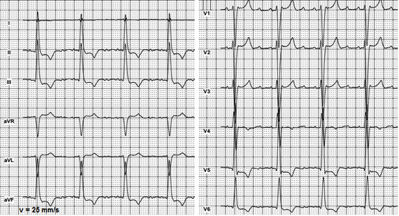
A pattern of irregular undulations of the base line in an electrocardiogram that is indicative of atrial fibrillation. Multifocal Atrial Tachycardia When multifocal atrial tachycardia occurs, multiple (non-SA) sites are firing impulses. Atrial flutter generates a defined pattern of atrial activity in the EKG with a "sawtooth" pattern in leads II, III, AVF without a defined isoelectric line between F waves (see figure 5a).
The waves of atrial flutter usually best seen in ECG leads 2, 3, and AVF. Electrodes are placed on your chest to record your heart's electrical signals, which cause your heart to beat. They are replaced by lower case "f" waves.
Therefore, each small 1-mm square represents 0.04. Although elicitable in a variety of muscles, it is best obtained in the small foot and hand muscles. And without the drawbacks and costly upkeep of asphalt shingles, natural wood shingles, and real slate roofing tiles.
The signals are shown as waves on an attached computer monitor or printer. The f waves result in an oscillating irregular baseline. The patient was agitated at the time of the original ECG resulting in an arte factual disturbance of the isoelectric line making the P waves difficult to discern.
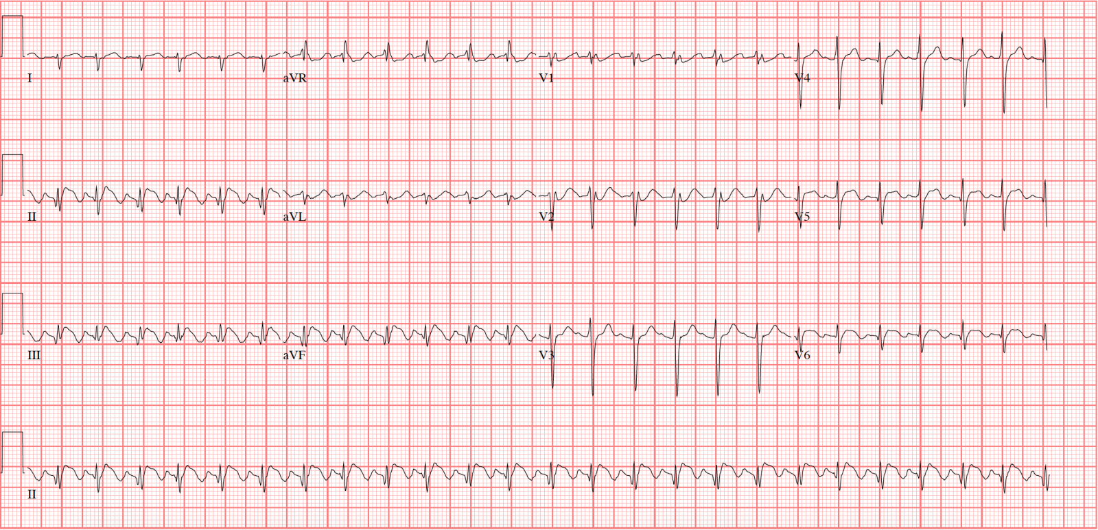
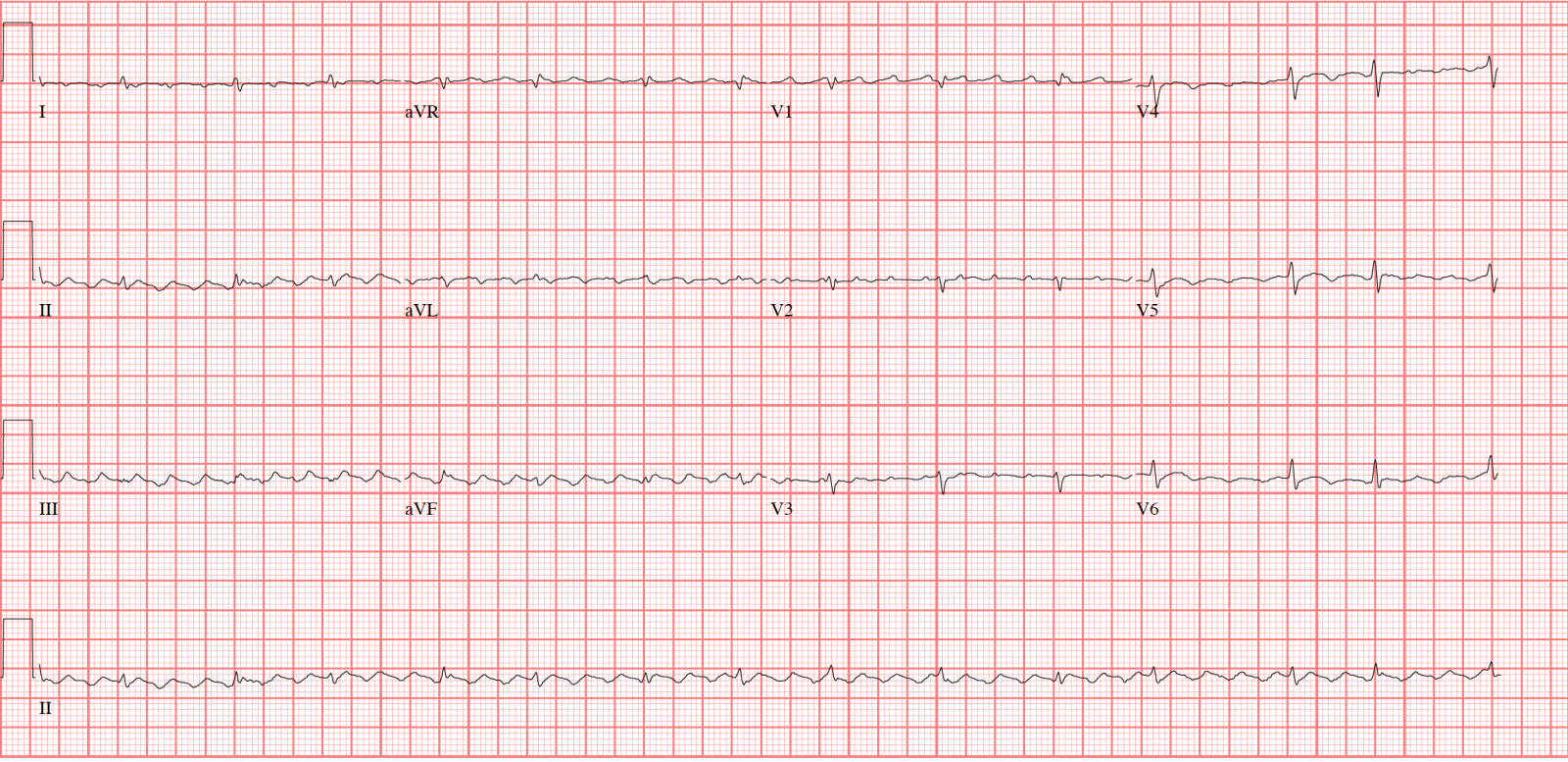
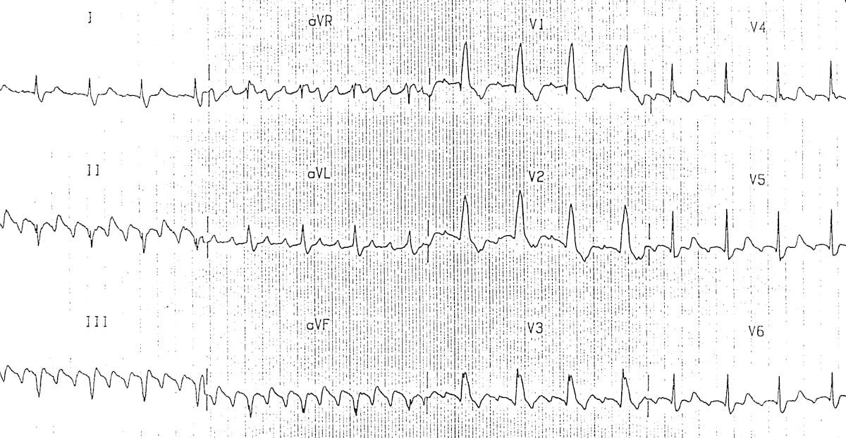
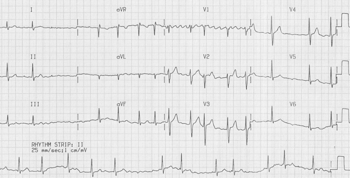
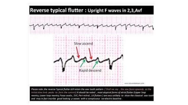
We know, classical Atrial flutter (Also referred to as typical /Common AF) records saw toothed F waves due to continuous atrial electrical activity across a macro- reentrant circuit within right atrium. Absent P waves on ECG:. ECG in atrial fibrillation The hallmark of atrial fibrillation is absence of P-waves and an irregularly irregular (i.e totally irregular) ventricular rate.
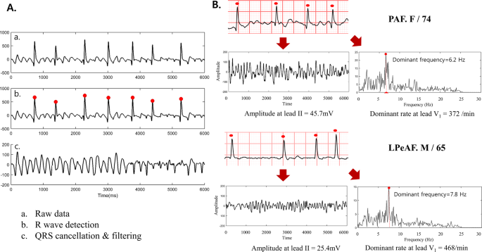
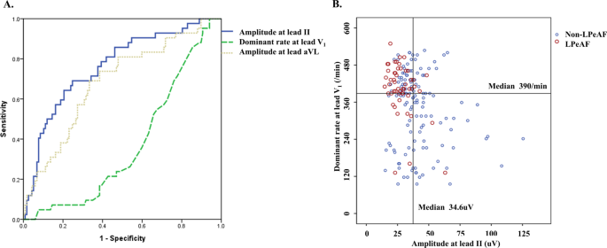
Differentiating anomalous left main coronary artery originating from the pulmonary artery in infants from myocarditis and dilated cardiomyopathy by electrocardiogram. Surface ECG f Wave Analysis at Initial Onset of Paroxysmal and Persistent Atrial Fibrillation. In neuroscience, an F wave is one of several motor responses which may follow the direct motor response evoked by electrical stimulation of peripheral motor or mixed nerves.
The F-waves of flutter can take a variety of morphologies, but most often the bulk of the wave is negative in the inferior leads and upright in V1. Aldose reductase inhibition ameliorates pupillary light reflex and F-wave latency in patients with mild diabetic neuropathy. F-waves are the second of two late voltage changes observed after stimulation is applied to the skin surface above the distal region of a nerve, in addition to the H-reflex which is a muscle reaction in response to electrical stimulation of innervating sensory fibers.
Representing ventricular repolarization, T waves are located after the QRS complex on an EKG. Also, as the atrial rate slows with the use of medications, there is a loss of F-wave amplitude and the morphology can become incredibly subtle. Small ‘septal’ Q waves are typically seen in the left-sided leads (I, aVL, V5 and V6).
The ECG findings of a pathologic Q wave include a Q wave duration of > 40 milliseconds (one small box) or size > 25% of the QRS complex amplitude. P waves are absent. The next area you see is a big spike.
The absence of a P wave located on an electrocardiogram. EKG tracings will show tightly spaced waves or saw-tooth waveforms (F-waves). Though this saw tooth pattern is easily recognised , it’s often difficult to say whether the saw is facing upwards or downwards ?.
As part of the QRS complex, an R wave is an important indicator of cardiac health.
Ppt Atrial Flutter And Atrial Fibrillation Powerpoint Presentation Free Download Id
Ecg In Aflutter

Atrial Fibrillation Af Atriyal Fibrilasyon Ekg Ecg Ankara Kardiyoloji Kalp Hastaliklari Mete Alpaslan Doktorekg Com
F Waves Ekg のギャラリー
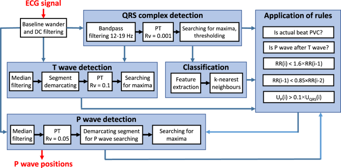
Advanced P Wave Detection In Ecg Signals During Pathology Evaluation In Different Arrhythmia Contexts Scientific Reports

F Waves In Ecg Dr S Venkatesan Md
Nursing Management Dysrhythmias Nurse Key
Atrial Flutter Classification Causes Ecg Diagnosis Management Ecg Echo
Atrial Flutter Litfl Medical Blog Ecg Library Diagnosis
Atrial Flutter Classification Causes Ecg Diagnosis Management Ecg Echo
11 Ecg Patterns Of Supraventricular Arrhythmias Thoracic Key

Ekg Strips Flashcards Quizlet

A Representative Example Of An Af Ecg Recording And Its Fiducial Download Scientific Diagram
The Physiological Basis Of The Ekg Ppt Download

Understanding The Ekg Signal Atrial Fibrillation Resources For Patients
Ecg In Aflutter

Ecg Amboss
Atrial Fibrillation Ecg Classification Causes Risk Factors Management Ecg Echo
Atrial Flutter And Atrial Fibrillation

Separating Atrial Flutter From Atrial Fibrillation With Apparent Electrocardiographic Organization Using Dominant And Narrow F Wave Spectra Sciencedirect
Interpretation Of Uncommon Ecg Findings In Patients With Atrial Flutter

Electrocardiography Radiology Key

Atrial Rhythms Lessons And Quiz

Atrial Fibrillation Complexity Parameters Derived From Surface Ecgs Predict Procedural Outcome And Long Term Follow Up Of Stepwise Catheter Ablation For Atrial Fibrillation Circulation Arrhythmia And Electrophysiology
10 Tips To Never Miss Atrial Flutter With 2 1 Conduction

Lesson Title Atrial Flutter
Atrial Fibrillation Litfl Medical Blog Ecg Library Diagnosis

Ecg A Pictorial Primer Medicine On Line Com
Atrial Flutter Litfl Medical Blog Ecg Library Diagnosis
1st Part Ecg Basics Indroduction And P Waves
1

Normal Ecg Waves Arryhthmias Ppt Video Online Download
Atrial Flutter Electrocardiogram Wikidoc
Q Tbn 3aand9gcqts24zy8 O4ata1y1l95dt16bmfklbcgfih0gkrhxza2ulzs Y Usqp Cau
Ppt Atrial Fibrillation Rhythm Powerpoint Presentation Free Download Id

Pin On Atrial Flutter
Atrial Fibrillation Wikipedia
Ecg Step By Step Not By Me

The 12 Rhythms Of Christmas Atrial Flutter Ems 12 Lead
11 Ecg Patterns Of Supraventricular Arrhythmias Thoracic Key

Electrolyte Abnormalities
Lesson Title Atrial Flutter

The Normal Ecg The Student Physiologist

Separating Atrial Flutter From Atrial Fibrillation With Apparent Electrocardiographic Organization Using Dominant And Narrow F Wave Spectra Sciencedirect
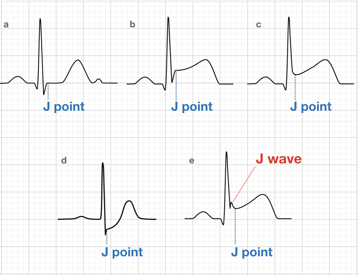
Osborn Wave J Wave Litfl Medical Blog Ecg Library Basics

Atrial Rhythms Lessons And Quiz
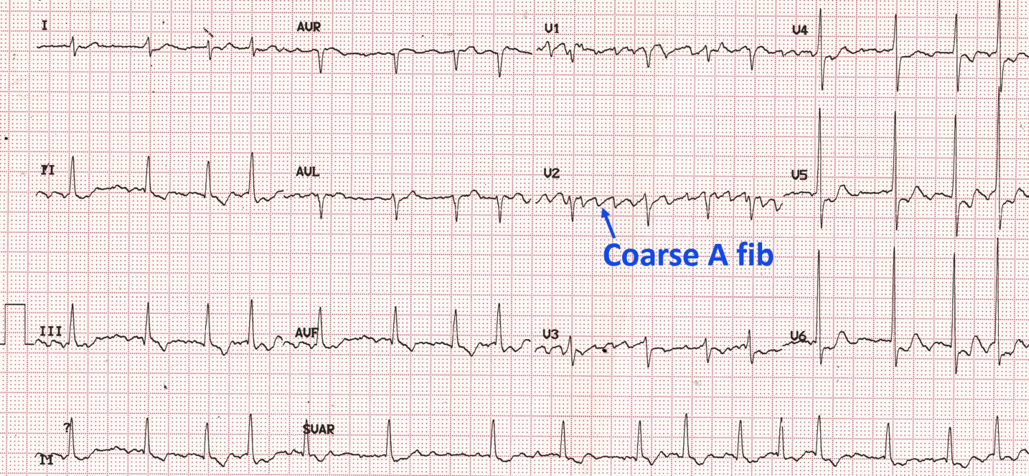
Coarse Atrial Fibrillation On Ecg All About Cardiovascular System And Disorders

Atrial Fibrillation Ecg Review Criteria And Examples Learntheheart Com
Atrial Flutter And Atrial Fibrillation
Early Differentiation Of Long Standing Persistent Atrial Fibrillation Using The Characteristics Of Fibrillatory Waves In Surface Ecg Multi Leads Scientific Reports
11 Ecg Patterns Of Supraventricular Arrhythmias Thoracic Key

Basic Dysrhythmia Kamlya Balgoon Ppt Video Online Download
Atrial Fibrillation Ecg Classification Causes Risk Factors Management Ecg Echo
Chapter 3 Supraventricular Rhythms Ii Atrial Fibrillation Ppt Download

Ecg Features Of Torsades De Pointes Tdp Torsades Depointes Tdp Diagnosis Cardiology Clinical

The 12 Rhythms Of Christmas Atrial Flutter Ems 12 Lead
Medicom Afib Detector
Q Tbn 3aand9gcq32uyamos1w 5 Fpabv3g 8vqhwbtrgy Pk7dny0aofl4auo8z Usqp Cau
Lesson Title Atrial Flutter

Lesson Title Atrial Flutter

Easy To Understand Different Between Knowledge Of Ecg Facebook

Ecg Amboss

The 12 Rhythms Of Christmas Atrial Flutter Ems 12 Lead
Atrial Flutter Litfl Medical Blog Ecg Library Diagnosis
Interpretation Of Uncommon Ecg Findings In Patients With Atrial Flutter

Ecg Primer For The Cath What Does A Tall R Wave In V1 Mean The Four Categories Approach Cath Lab Digest

7 Can T Miss Life Threatening Ecg Findings
Q Tbn 3aand9gcqd5hwdwpwg5ur3ji Zbas6f3i7 0qjfamk Gqtspz Fl9sddo Usqp Cau
Q Tbn 3aand9gcriu0a6yr6 Kpsblb1nf9evv3fhckzri Zbhq Usqp Cau

The 12 Rhythms Of Christmas Atrial Flutter Ems 12 Lead
Early Differentiation Of Long Standing Persistent Atrial Fibrillation Using The Characteristics Of Fibrillatory Waves In Surface Ecg Multi Leads Scientific Reports

Ecg A Pictorial Primer Medicine On Line Com

Ecg Amboss

The Extraction Of The F Waves And A Typical Example A Data Download Scientific Diagram

Conclusion To 68 Yof Cc Unresponsive On Kitchen Floor Osborn Waves Of Hypothermia Ems 12 Lead
Cardiac Arrhythmia Ppt Video Online Download

Atrial Flutter Wikipedia
Lesson Title Atrial Flutter
Solved What Is The Heart Rate 60 1 Unable To Determine Chegg Com
Atrial Flutter Ecg Acls Wiki

Atrial Fibrillation Topic Review Learntheheart Com

Ekg Strips Flashcards Quizlet
Atrial Fibrillation Ecg Classification Causes Risk Factors Management Ecg Echo
Atrial Flutter Litfl Medical Blog Ecg Library Diagnosis
Atrial Flutter And Atrial Fibrillation
Electrocardiogram Physiopedia
Atrial Fibrillation Ecg Classification Causes Risk Factors Management Ecg Echo
Co Grand Co Us Documentcenter View
Atrial Fibrillation Litfl Medical Blog Ecg Library Diagnosis

What Is The Difference Between Atrial Fibrillation A Fib Atrial Flutter A Flutter
Ecg In Aflutter

Epsilon Wave An Overview Sciencedirect Topics

Electrocardiography Cardiovascular Disorders Msd Manual Professional Edition

A Representative Example Of An Af Ecg Recording And Its Fiducial Download Scientific Diagram

Atrial Fibrillation Af Atriyal Fibrilasyon Ekg Ecg Ankara Kardiyoloji Kalp Hastaliklari Mete Alpaslan Doktorekg Com

Reverse Typical Clockwise Atrial Flutter On Ekg Inferior Leads Positive Waves
Co Grand Co Us Documentcenter View

Causes Of Cardiac Arrhythmias Ppt Video Online Download

Pdf Intracardiac Overdrive Pacing As A Treatment Of Atrial Flutter In A Horse

Delineation Of The F Waves For Typical 4 S Segments Corresponding To Download Scientific Diagram
The P Wave Ecg Basics Medschool

What Is The Difference Between Atrial Fibrillation A Fib Atrial Flutter A Flutter

Atrial Fibrillation Af Atriyal Fibrilasyon Ekg Ecg Ankara Kardiyoloji Kalp Hastaliklari Mete Alpaslan Doktorekg Com
Lesson Title Atrial Flutter

The 12 Rhythms Of Christmas Atrial Flutter Ems 12 Lead


